![]() Make Appointments 24/7, 365 days a year.
Make Appointments 24/7, 365 days a year. ![]() 302-731-2888
302-731-2888
![]() Make Appointments 24/7, 365 days a year.
Make Appointments 24/7, 365 days a year. ![]() 302-731-2888
302-731-2888
Matthew A. Handling, M.D.
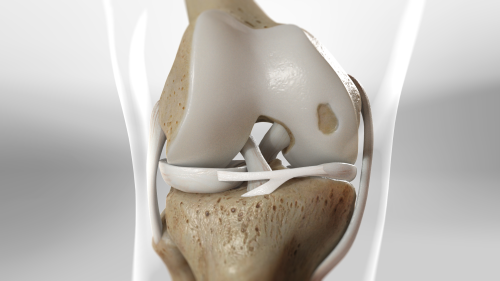
Cartilage is the strong and smooth substance that makes up the gliding surfaces in our joints. It is made up of cells call chondrocytes. Cartilage injuries can occur in any joint but are most-commonly found in weight bearing joints like the knee and ankle. The goal of treatment for any cartilage defect or injury is preserving the person’s normal cartilage to prevent future osteoarthritis. Unfortunately, our bodies do not have the ability to heal or repair a cartilage defect with normal cartilage cells. Luckily, there are many different types of surgical options for cartilage injuries.
The symptoms that people feel for focal cartilage defects include pain, swelling, stiffness, and locking or catching, which can interfere with activities. Traumatic injuries like a knee-cap dislocation or an ACL tear can cause a cartilage defect, but also repetitive overload can produce cartilage damage. An OCD or Osteochondritis dissecans is a specific type of cartilage lesion that occurs in children. Most cartilage defects are diagnosed on an MRI study. This helps determine the size and location of the defect which aids in surgical planning. An MRI will also pick up any other injuries in a knee such as a ligament tear (ACL) or meniscus tear.
Surgical treatment options include:
Cartilage injuries can be a challenging problem for patients. The above treatment options will be individualized to each patient based on location, size, and depth of the injury to the cartilage. If you have cartilage injury or would like a second opinion, then contact Dr. Matthew Handling at First State Orthopaedics and make an appointment today.
* indicates a required field.